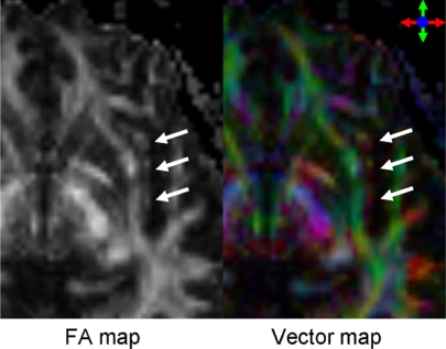
I read the article by Saur et al. (1) with interest. The authors state that they succeeded in in vivo depiction of the dual-stream model for language processing, which has been recently suggested by Hickok et al. (2, 3). These fibers are known to course through the extreme capsule (EmC), which is a fiber bundle that is located lateral to the external capsule (EC), divided by a thin layer of gray matter known as the claustrum. The authors of this article have combined functional MRI and probabilistic tractography to show that higher-level language comprehension is mediated by a ventral pathway, running in an anteroposterior direction within the EmC. The data seemed convincing until I tried to reproduce their results using my own dataset obtained by using a similar spatial resolution (2 × 2 × 2 mm) (4). By carefully looking into the color-coded vector map (Fig. 1), I immediately noticed that the fiber direction of the EmC is not anteroposterior but rather transverse or superoinferior in the transaxial plane (indicated by the deep purple color). On the other hand, the EC had fibers running in an anteroposterior direction (shown in green). It thus seems that their probabilistic tractography has erroneously picked the EC (rather than the EmC) to elucidate an imaginary fiber tract connecting the temporal lobe and the ventrolateral prefrontal cortex. As a clinician, I have been using this tool (tractography) with the greatest caution, because all that we see on these intriguing images may not precisely reflect the reality (5). I think that the results of Saur et al. (1) represent another example of such false positive/negative results.
Fig. 1.
The fractional anisotropy (FA) and color-coded vector maps at the level of the basal ganglia. The vector map shows the direction of the local fibers, represented by red (left to right), green (anterior to posterior), and blue (superior to inferior). The location of the EmC is indicated by the arrows.
Footnotes
The author declares no conflict of interest.
References
- 1.Saur D, et al. Ventral and dorsal pathways for language. Proc Natl Acad Sci USA. 2008;105:18035–18040. doi: 10.1073/pnas.0805234105. [DOI] [PMC free article] [PubMed] [Google Scholar]
- 2.Hickok G, Poeppel D. Dorsal and ventral streams: A framework for understanding aspects of the functional anatomy of language. Cognition. 2004;92:67–99. doi: 10.1016/j.cognition.2003.10.011. [DOI] [PubMed] [Google Scholar]
- 3.Hickok G, Poeppel D. The cortical organization of speech processing. Nat Rev Neurosci. 2007;8:393–402. doi: 10.1038/nrn2113. [DOI] [PubMed] [Google Scholar]
- 4.Yamada K, et al. Somatotopic organization of thalamocortical projection fibers as assessed with MR tractography. Radiology. 2007;242:840–845. doi: 10.1148/radiol.2423060297. [DOI] [PubMed] [Google Scholar]
- 5.Kinoshita M, et al. Fiber-tracking does not accurately estimate size of fiber bundle in pathological condition: Initial neurosurgical experience using neuronavigation and subcortical white matter stimulation. NeuroImage. 2005;25:424–429. doi: 10.1016/j.neuroimage.2004.07.076. [DOI] [PubMed] [Google Scholar]


