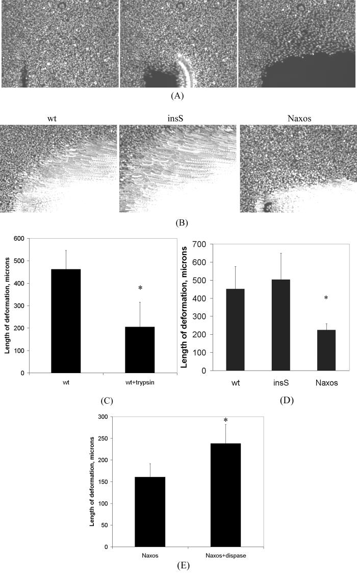
Figure 6.
Deform-drag experiments performed on confluent cell monolayers. (A) Representative images illustrating the principal of the deform-drag method in a culture of cells expressing wildtype wt) plakoglobin (10x magnification). A glass rod was placed near a corner of the field of view (lower left region of the left image), where a small wound was created. The cells had been labeled with Hoechst to visualize nuclei in the live state. The rod was then dragged across the field using a motorized micromanipulator (from left to right), resulting in removal of cells in the path of the rod (see movies in supplemental files). The resulting wound can be seen in the center and right images. During active wounding, cells adjacent to the region of wounding may be pulled (deformed) along as cells in the rod's path are removed. The extent of deformation of adjacent cells is a function of the strength of cell-cell adhesiveness. (B) By merging the frames acquired during dragging, the motion of nuclei in adjacent cells from cultures expressing wildtype or mutant plakoglobin can be visualized and quantified. The extent of motion in the vertical direction (e.g., perpendicular to the direction of the wound) can be used as a measure of the strength of cell-cell adhesiveness, assuming comparable cell-matrix adhesiveness. (C) A validation experiment showing that brief exposure to trypsin of cells expressing wildtype plakoglobin significantly decreases drag-deformation (* p<0.005). (D) Group data showing that cells expressing the Naxos mutation, but not the insS mutation, exhibit markedly diminished cell-cell adhesiveness compared to wildtype cells (n = 6 for each group; * p<0.05 compared to wildtype and insS). (E) A second validation experiment showing that brief dispase treatment significantly increases drag-deformation in cells expressing Naxos plakoglobin (* p<0.005), suggesting that the balance of cell-matrix and cell-cell adhesion has shifted toward the latter.

