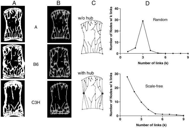
Fig. 1.

Conversion of 2D vertebral images into network maps. (A) Two-dimensional coronal sections were (B) skeletonized and nodes were identified. (C) The skeleton graphs were converted into network maps by considering the cortical surface as one highly connected node or hub (large circles). (D) Graphical representation of random (no hub) and scale-free (cortical hub) networks.
