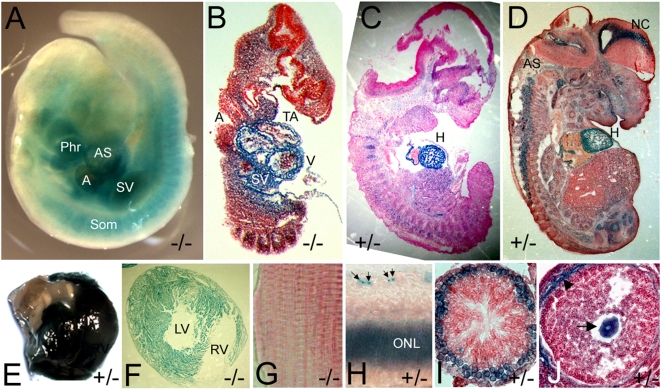
Figure 2. The expression pattern of the Asxl2 gene trap in developing embryos and adult organs.
Developing embryos and postnatal organs were stained with X-gal. (A) A whole-mount E9.5 embryo. (B–D) Cryo-sections of whole-mount stained E10.5 (B), E12.5 (C) and E14.5 (D) embryos. (E) Top view of a whole-mount stained postnatal heart showing strong X-gal staining in the heart but not in the aorta. (F) Cryo-section of a whole-mount stained postnatal heart. (G) Cryo-section of a piece of whole-mount stained skeletal muscle. The skeletal muscle in (G) and the heart in (F) were taken from the same animal and stained together. There was no detectable X-gal activity in the skeletal muscle. (H) Cryo-section of the retina in a whole-mount stained eye. Arrows point to stained cells in the ganglion cell layer. (I) Cross-section of a seminiferous tubule in a whole-mount stained testis showing expression in the primary spermatocytes. (J) Cross-section of an early tertiary follicle in a whole-mount stained ovary, showing expression in the oocyte (arrow). The blue color around the periphery of the follicle (arrowhead) was non-specific and also detected in wild-type samples. Phr: 1st pharyngeal arch; A: common atrial chamber; AS: aorta sac; SV: sinus venosus; Som: somites; V: common ventricular chamber; TA: truncus arteriosus; H: heart; AS: axial skeleton; NC: neocortex; RV: right ventricle; LV: left ventricle; ONL: outer nuclear layer.

