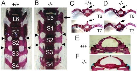
Figure 4. The loss of Asxl2 disrupts axial skeleton patterning.
(A, B) Ventral view of the sixth lumbar vertebra (L6) and the sacral vertebrae 1–4 (S1–4). (A) A wild-type skeleton. The wing-shaped lateral processes of S1, S2 and S3 fuse at the tip (arrowheads). The lateral processes of S4 do not fuse with those of S3. (B) A mutant skeleton. The lateral processes of S1, S2 and S3 fail to fuse. Arrowheads point to the sites at which fusion should happen. The lateral processes of mutant S2 resemble those of wild-type S3; those of mutant S3 resemble those of wild-type S4. One of the lateral processes of mutant L6 resembles that of S1 (arrow). (C, D) Caudal view of isolated T6 and T7 vertebrae, showing anterior transformation in the mutant. The wild-type T6 and T7 differ in the length of the neural spine (arrows) and the arch of the attached ribs (C). The mutant T7 resembles T6 in both the neural spine and the arch of ribs. (E, F) Caudal view of the first thoracic vertebrae (T1) in a heterozygous skeleton (E) and a mutant skeleton (F). The mutant T1 is split at the dorsal midline (asterisk).

