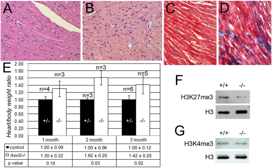
Figure 5. Cardiac phenotypes of Asxl2−/− animals.
(A, B) H&E stainings of heart sections. The myocardial fibers, composed of cardiomyocytes, are well organized in the wild-type heart (A). In contrast, the heart of a moribund 5-month-old Asxl2−/− mouse shows cardiomyocyte disarray (B). (C, D) Examination of fibrosis in heart sections by Mason's trichrome (MTC) staining. Blue staining marks regions of fibrosis. The mutant (D) but not the wild-type (C) heart displays extensive interstitial fibrosis. (E) The heart/body weight ratios of mutant and control animals at 1 month, 2 months and 3 months after birth. The table below the chart shows the average ratio + standard deviation for each genotype and the p value for the time point. Error bars in the chart represent standard deviations. (F, G) Western blot analysis of the levels of H3K27me3 and H3K4me3 in wild-type and mutant hearts. As a control for equal loading, the blots were stripped and re-probed with an antibody against pan histone H3. The mutant heart has decreased level of H3K27me3 (F). There is no obvious change in the level of H3K4me3 (G).

