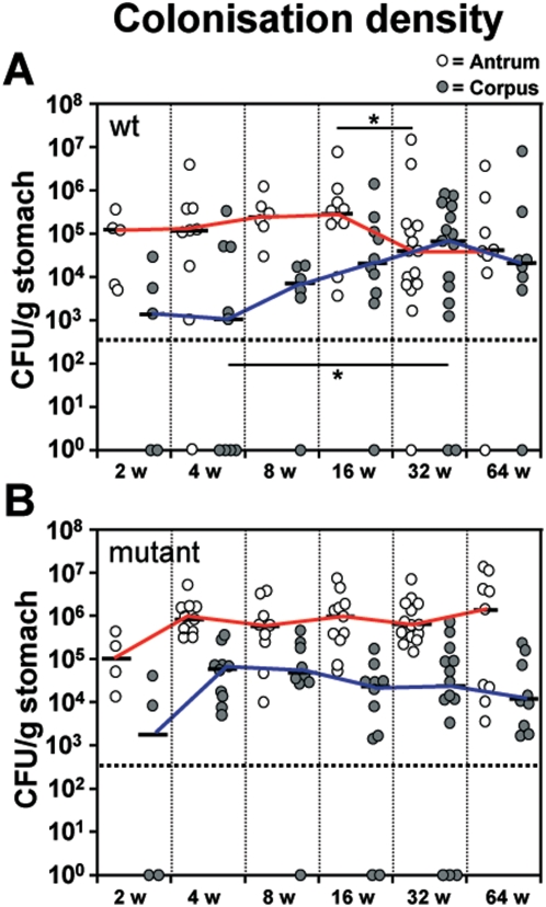
Figure 1. Increased Helicobacter pylori B128 WT colonization density shown in corpus mucosa over the time course experiment.
Colonization density of antral (white circles) and corpus (gray circles) mucosa in orally challenged gerbils with H. pylori B128 WT (A) and B128ΔcagY (B) isogenic mutant strain after 2, 4, 8, 16, 32, and 64 weeks of infection. The interpolated lines connect the medians of the respective time-points. The detection limit was <5×10² colony-forming units (CFU) per gram of stomach (horizontal dotted line). Gastric tissue specimens without H.pylori reisolation are shown as null. (*p<0.05).

