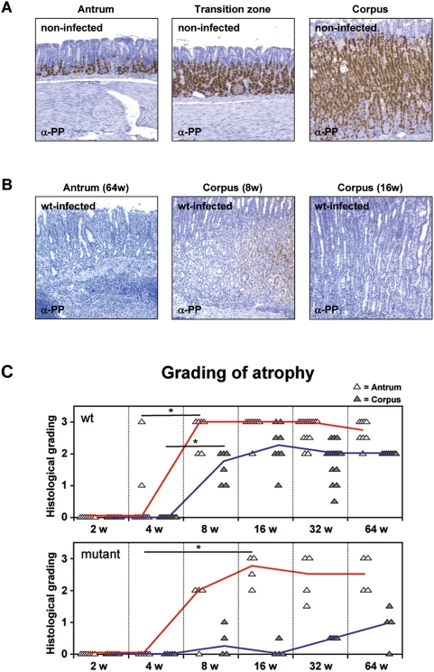
Figure 4. Distribution of parietal cells analyzed by immunohistochemical staining.
(A) Parietal cells were detected with anti-proton pump antibody in antral tissue (left panel), transition zone (middle panel), and corpus tissue (right panel) of non-infected Mongolian gerbils (original magnification 10×). (B) Immunohistochemical staining with anti-proton pump antibody detecting complete parietal cell atrophy in antral tissue at 64 weeks (left panel) and beginning at 8 weeks (middle panel) as well as complete parietal cell atrophy in corpus tissue at 16 weeks (right panel) of H. pylori B128 WT-infected gerbils. (C) Histological parietal cell atrophy grading of antral (white triangles) and corpus (gray triangles) mucosa infected with H. pylori B128 WT (upper panel) and B128ΔcagY isogenic mutant strain (bottom panel) over time. (*p<0.05).

