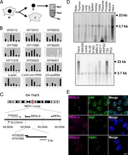
Fig. 5.
MENε/β ncRNAs and the paraspeckle marker proteins cooperate to organize the paraspeckle structure. (A) The MENε/β levels were quantified by qRT-PCR upon RNA interference (RNAi) of each paraspeckle protein. The relative abundance (100% in control cells treated with a control oligonucleotide) is shown in the graph. Two siRNAs were used for each paraspeckle protein. Numbers below the graphs correspond to the primer pairs for qPCR, as in Fig. 2A. Values represent means ± SD. *, P < 0.01. (B) The effect of RNAi on the paraspeckle structure was monitored by FISH-IF. Cells were treated with the siRNA indicated above each panel for 48 h and were then probed with MENε/β probe (green) in combination with an antibody to one of the three paraspeckle proteins (magenta: p54, PSF, or PSP1, top to bottom). Arrows point to the remnant MENε/β puncta with paraspeckle proteins. Open triangles point out imperfect MENε/β puncta lacking at least 1 paraspeckle protein. (Scale bar: 10 μm.) (C) Immunoprecipitation (IP) of MENε/β with antibodies against paraspeckle proteins. Flag-tagged p54 was used instead of endogenous p54 for p54-IP. Numbers below the graphs correspond to those in Fig. 2A. Values represent means ± SD. *, P < 0.01. (D) The paraspeckle protein p54 directly interacts with MENβ ncRNA. Intact HeLa cells transfected either with Flag-p54 or a control plasmid (FLAG-pcDNA) were irradiated with 254-nm UV light to induce cross-links between RNA and interacting proteins in vivo (+UV). Cell extracts were prepared under strong denaturing conditions and subsequently subjected to IP with the αFlag antibody. The relative IP efficiencies (the practical IP efficiency detected by primer #4 was defined as 100%) are shown, because IP efficiency in each of 3 independent experiments was variable (1.23%, 2.70%, and 11.5%). The large fluctuation in IP efficiency may be due to the efficiency of UV cross-linking. Values represent means ± SD. *, P < 0.05. (E) Possible interaction between MENε/β ncRNAs and 3 known paraspeckle proteins that underlie paraspeckle organization. The protein–protein interactions are depicted according to previous reports (12, 21).

