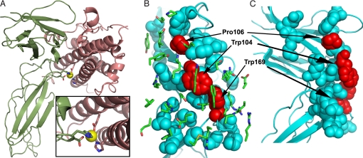
Fig. 3.
hGH–hGHR complex structures. (A) Ribbon structure of the hGH(salmon)–hPRLR(green) complex highlighting the interface Zn2+-binding site. The four Zn2+ ligands include hGH residues His-18 and Glu-174 and hPRLR residues Asp-187 and His-188. (B) View of the hGHR interface residues (blue and red spheres) illustrating the location of the transition state hot-spot residues (Trp-104, Pro-106, and Trp-169; shaded red). The Trp residues represent the knobs that fit into the hGH interface hole. The Site 1 hGH interface residues (green sticks) are displayed in the foreground. (C) Side view of the hGHR interface displayed in B. Figures were generated with PyMOl (35).

