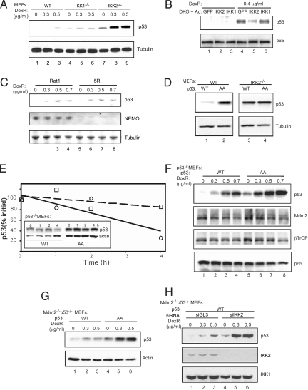
Fig. 2.
Phosphorylation by IKK2 destabilizes p53. Primary WT, IKK1−/−, and IKK2−/− MEF cells (A); IKK1/2−/− DKO MEF cells infected with recombinant adenovirus expressing GFP, IKK2, and IKK1 (B); Rat1 and 5R rat fibroblast cells (C); Primary WT and IKK2−/− MEF cells stably infected with Flag-p53 WT or AA lentivirus (D); p53−/− MEF cells stably infected with Flag-p53 WT or AA lentivirus (F); Mdm2−/−/p53−/− MEF cells infected with Flag-p53 WT or AA lentivirus (G); Mdm2−/−/p53−/− MEF cells expressing p53 WT infected with siGL3 or siIKK2 lentivirus (H) were treated with 0.4 μg mL−1 or increasing amounts of doxorubicin for 18 h. Cell lysates were immunoblotted with indicated antibodies. (E) Doxorubicin-treated p53−/− MEF cells stably expressing recombinant Flag-p53 WT or AA protein were collected at the indicated time points after addition of cyclohexamide (CHX) for 0, 1, 2, and 4 h. The data shown in the inset were quantified by densitometry. The dashed line represents p53 AA, and the solid line represents p53 WT.

