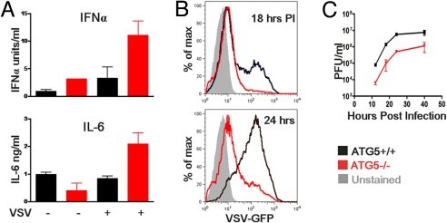
Fig. 1.
Atg5-deficient MEFs show increased cytokine production to Poly I:C and VSV stimulation and are resistant to infection. WT, and Atg5−/− MEFs were incubated with VSV-GFP (at a multiplicity of infection of 4) or transfected with 1 μg/mL Poly I:C. Twelve hours later, IFNα and IL-6 production was assessed by ELISA (A). WT and Atg5−/− MEFs were infected with VSV-GFP (at a multiplicity of infection of 1) and the levels of infection were determined by measuring GFP expression by FACS at 18 and 24 h after infection (B) and by measuring viral titers in the supernatants by plaque assay at the indicated time points (C). Results are representative of 6 separate experiments.

