Fig. 5.
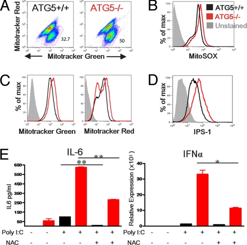
Atg5-deficient primary macrophages exhibit amplified RLR signaling. Neonatal liver macrophages were generated from Atg5+/+ or Atg5−/− pups. Cells were stained with 100 nM MitoTracker Green and 100 nM MitoTracker Red as in Fig. 2, and both dot plots (A) and histograms (C) of FACS analysis are depicted. Additionally, cells were labeled with MitoSOX as in Fig. 4 (B) or with antibody to IPS-1 as in Fig. 3. (D) Primary macrophages were transfected with 10 μg/mL Poly I:C in the presence or absence of NAC, and 12 h later, the levels of IL-6 were assessed by ELISA and the levels of IFNα were assessed by RT quantitative PCR (E). *P < 0.05, **P < 0.005. Results are representative of 2 similar experiments.

