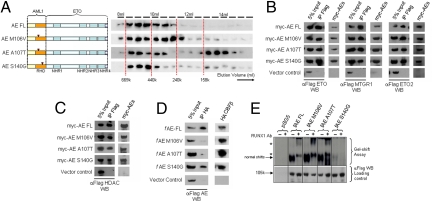
Fig. 1.
Identification and biochemical characterization of AE point mutants defective in CBFβ interaction. (A) Left: Gel filtration analysis on AE point mutants. Right: Western blot using α-Flag antibody on protein fractions collected from indicated elution volumes; dotted lines indicate the positions of the corresponding size standards. (B–D) Coimmunoprecipitation assay between (B) myc-tagged AE constructs and Flag-tagged ETO family members, (C) myc-tagged AE constructs and Flag-tagged HDAC1, or (D) Flag-tagged AE constructs and HA-tagged CBFβ. Antibodies used for immunoprecipitation (IP) and Western blot (WB) are indicated. (E) Top, Gel shift assay using lysates from 293 cells transfected with indicated constructs together with P-end-labeled CD11a probe in the absence or presence (for supershift) of AML1 antibody. Asterisks indicate supershifted bands. Bottom, Western blot of cell lysates used for gel shift experiment using α-Flag antibody.

