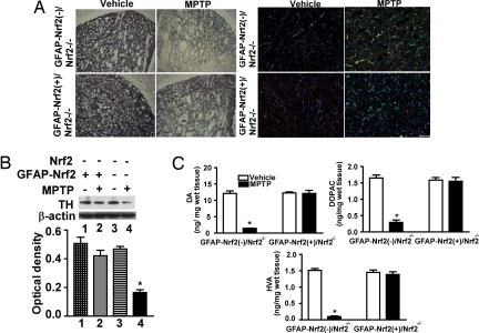
Fig. 6.
Effect of astrocyte-specific Nrf2 overexpression on MPTP toxicity in a Nrf2−/− background. (A) (Left) Immunohistochemical staining for TH in the STR of GFAP-Nrf2(−)/Nrf2−/− and GFAP-Nrf2(+)/Nrf2−/− mice after vehicle or 30 mg/kg MPTP treatment. (Right) Staining for GFAP (green) and Iba-1 protein (red) in the STR of GFAP-Nrf2(−)/Nrf2−/− and GFAP-Nrf2(+)/Nrf2−/− mice after vehicle or MPTP treatment. Fig. S6 is an enlargement of A. (B) (Upper) Representative Western blots of TH and β-actin. (Lower) TH signal was normalized to β-actin signal for protein loading control, and the bar shows quantification of the TH Western blots (n = 8–10). (C) The amount of DA, DOPAC, and HVA in the STR of GFAP-Nrf2(−)/Nrf2−/− and GFAP-Nrf2(+)/Nrf2−/− mice after vehicle or MPTP treatment (n = 8–10). (Scale bar, 50 μm.) *, P < 0.05 compared with the corresponding vehicle-treated group).

