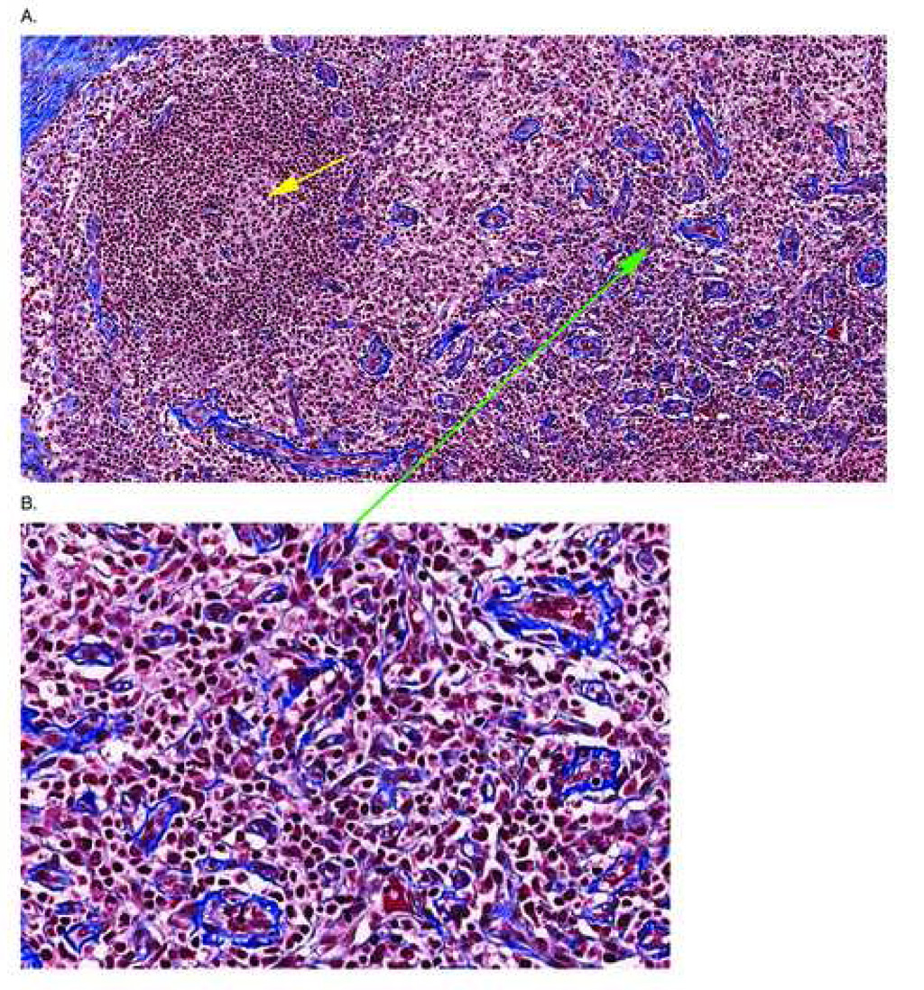
Figure 2. Representative Image of a T Cell Zone Stained With Trichrome.
A section of lymph node from a patient with AIDS is stained with trichrome to reveal collagen deposition in the tissues. In Panel A the T cell zone is evident by the numerous HEV’s (green arrow) showing thickened walls and there is significant deposition of collagen in the tissues of the T cell zone surrounding the HEV’s (more evident in the enlargement of the area around the green arrow shown in Panel B). The yellow arrow shows an involuted, burned out follicle that is characteristic in lymphatic tissues of individuals with advanced disease

