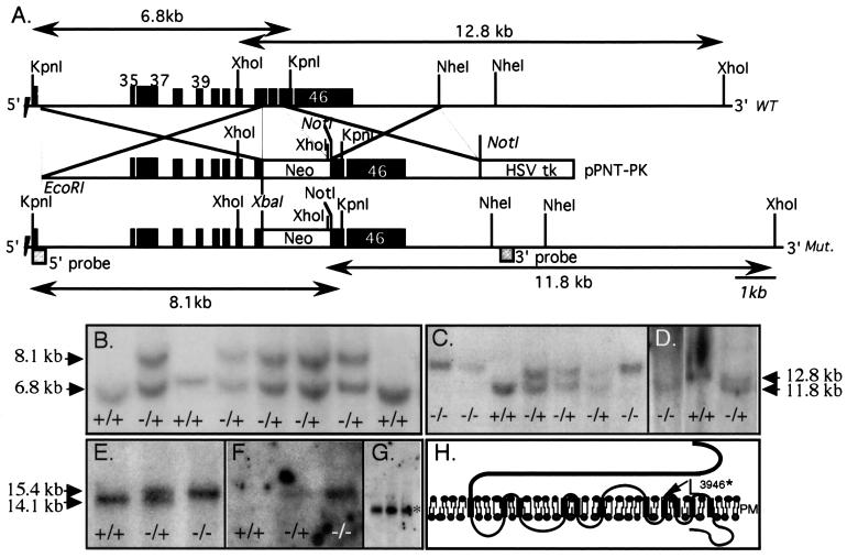
Figure 1.
Targeted disruption of mouse Pkd1. (A) The genomic segment of WT and recombinant murine Pkd1. Exons (black boxes) are numbered. The position of the recombinations, the expected sizes of WT and mutant (Mut.) fragments, and the position of the external 5′ and 3′ probes are indicated. (B) Southern blots of genomic DNA extracted from ES clones, KpnI-restricted, and probed with the 5′ external probe. A recombinant mutant fragment (8.1 kb) replaces the normal allele (6.8 kb) in several clones. DNA from clone #282 and #130 is shown in lanes 2 and 6. (C and D) Germ-line transmission of the mutant allele. Yolk sac DNA obtained from the offspring of F1 intercrosses was extracted, digested with KpnI or XhoI, and probed, respectively, with the 5′ (C) or 3′ (TSC2 exon 33, 430 bp, D) probes. Fragment size is indicated by arrows. Southern blots of DNA from clone #282 (lanes 1–4 in C and 1–3 in D) and #130 (lanes 5–7, C) F2 embryos is shown. +/+, +/−, and −/− indicate WT, Pkd1+/L, and Pkd1L/L, respectively. (E--G) Northern blot analysis. The mutant allele (15.4 kb) is found in Pkd1L/L (lane 3) and Pkd1+/L (lane 2) but not WT animals, who express only the WT mRNA of 14.1 kb (lane 1). The mutant but not the WT allele hybridized with the neo probe (F). (G) The normal-sized mRNA for mouse TSC2 (5.6 kb, *) in Pkd1L/L embryos. (H) Primary structure of polycystin 1 and the site of the predicted truncation (arrow). Mutant mouse polycystin 1 is expected to terminate after L3946 (equivalent to L3955 in human).

