Figure 4.
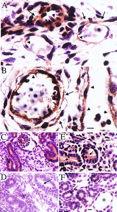
Expression of polycystin 1 in WT and Pkd1L/L E14.5 mouse embryos. Polycystin 1 is detected in endothelial cells (arrowheads in A and B) as well as in vascular smooth muscle cells (arrows in A and B) by using the anti-LRR antibody. Polycystin 1 expression (horseradish peroxidase reaction product) is similar in small vessels in the region of the hind limb in WT (A) and Pkd1L/L mutant (B) embryos. In fetal kidneys, polycystin 1 expression is observed mainly on the apical (arrows in C and E) surfaces of WT and Pkd1L/L renal tubules. No staining of renal tubules (arrows) from either WT or Pkd1L/L embryos is observed in the presence of a 20-fold excess of the LRR fusion protein (D and F, respectively). * in F points to a tubular cyst. (Scale bars = 10 μm.)

