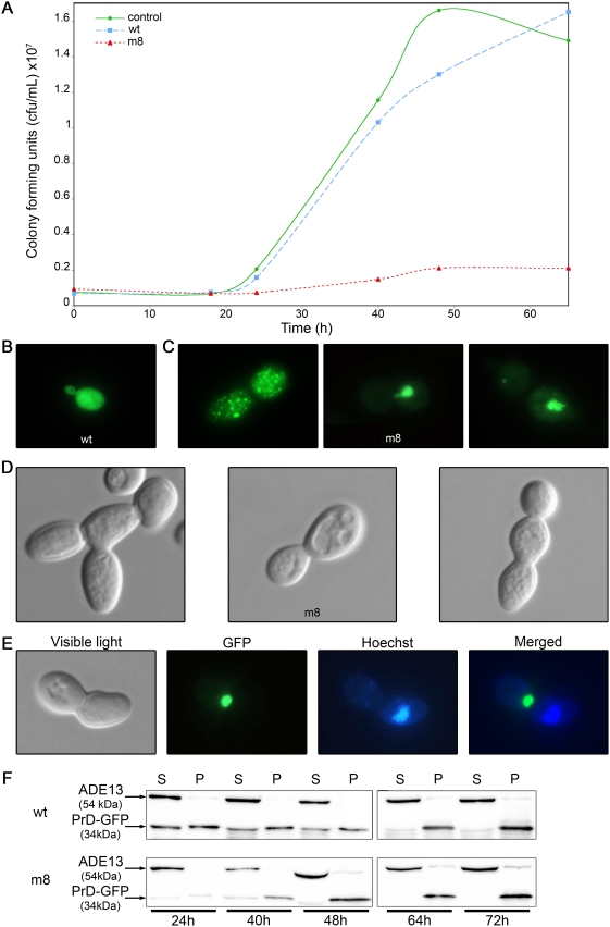
Figure 3. The m8 HET-s(PrD)-GFP mutant impairs cell viability.
(A) Toxicity of m8 expression demonstrated by a viability assay. The strain BY4742 was transformed with a plasmid expressing either wt or m8 HET-s(PrD)-GFP or a control plasmid. Liquid galactose medium was then inoculated and grown for 18 to 65 hours before being plated onto glucose solid medium. Colonies were numbered, and this value was then converted to colony forming units (cfu/mL). (B) Cells expressing wild-type HET-s(PrD)-GFP were examined with a fluorescence microscope after 24 h of growth in liquid galactose medium. (C) Cells expressing m8 HET-s(PrD)-GFP were observed using a fluorescence microscope after 24 h of growth in liquid galactose medium. (D) Cells expressing m8 HET-s(PrD)-GFP were observed with a light microscope after 24 h of growth in liquid galactose medium, using a Nomarski contrast filter. (E) Cells expressing m8 HET-s(PrD)-GFP were examined using both a light and a fluorescence microscope after 24 h of growth in liquid galactose medium. GFP fluorescence (green) and Hoechst DNA staining (blue) of an isolated dividing cell are shown. A GFP/DNA merged image was made using Adobe Photoshop. (F) Distribution of the GFP species between pellet (P) and supernatant (S). An aliquot of the culture used in the previous cfu assay was pipetted out after 24 to 72 hours. Total cell extracts underwent ultracentrifugation for 30 min; supernatant and pellet were run on a 12% SDS-PAGE gel, and were transferred onto a nitrocellulose membrane and exposed to anti-GFP antibodies.

