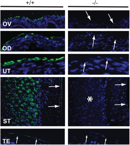
Figure 3. Immunohistochemistry of adult Muc16 wild-type (+/+) and homozygous mutant (−/−) tissues.
MUC16 protein (arrows) was absent in Muc16 −/− tissues. Asterisk indicates the loss of MUC16 protein in the chief cells in the stomach. OD, oviduct; OV, ovary; ST, stomach; TE, testis; UT, uterus. Scale bar = 50 µm.

