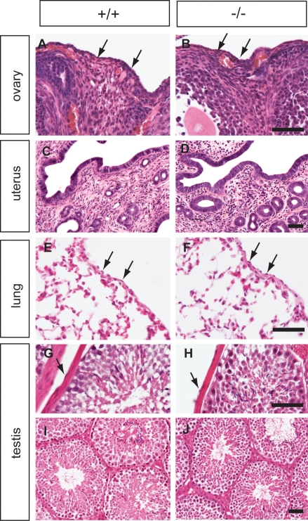
Figure 5. Histology of Muc16 wild type (+/+) and homozygous mutant (−/−) tissues.
The ovary (A, B), uterus (C, D), lung (E, F), and testis (G–J) from wild-type (left panel) and homozygous mutant animals (right panel) were analyzed by H&E staining. Tissues were collected at 3 months (A–B, E–J) and 1 year of age (C, D). Arrowheads indicate mesothelial cells. Scale bars = 50 µm.

