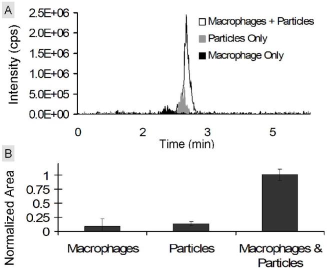
Figure 7.

Detection of compound 2 in vitro. RAW macrophages were cultured with polymer 1 particles for 48 hours. Polymer 1 particles incubated in medium and macrophages alone served as controls. (A) Representative extracted chromatograms for compound 2 of RAW macrophages incubated with particles (white), macrophages alone (black), or polymer 1 particles incubated in medium alone (gray). (B) Average areas calculated from the extracted chromatograms of compound 2 in each sample. For each sample, the area for compound 2 was divided by the area of an internal standard (compound 6) and normalized with respect to the maximum average area (macrophage & particle sample).
