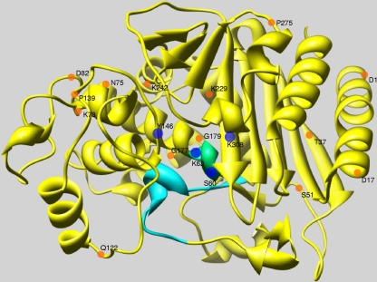
FIG. 3.
Theoretical model of the crystallographic structure of the ADC-12 AmpC β-lactamase. Amino acids belonging to the main β-lactamase domains (SVSK, YSN, and KTG) are represented in the figure as blue circles (60-63SXXK, Y146, and K308). The SVSK domain is also shown in green. The Ω-loop is shown in turquoise. Residues at positions 1, 17, 37, 51, 75, 78, 82, 122, 139, 177, 229, 242, and 275 (Fig. 1), which were replaced in four or more ADC-type enzymes (relative to ADC-1), are represented by orange circles (starting at position +1 of the mature protein with D). The diagram was drawn with the USCF Chimera software package.

