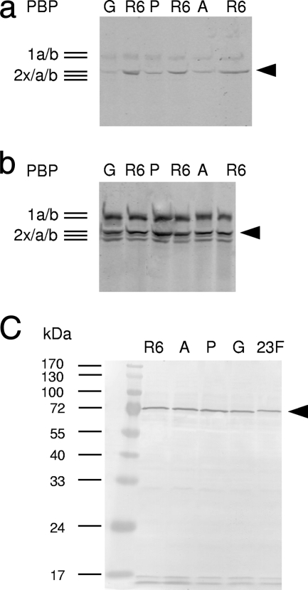
FIG. 1.
PBP profiles of PBP2x mutants. Cell lysates were incubated for 2 min at 20°C. (a and b) BocillinFL at 0.01 μM (a) or 0.3 μM (b) final concentration. Bocillin-PBP complexes were visualized by fluorography after separation on SDS-polyacrylamide gels. (c) Detection of PBP2x after Western blotting of cell lysates and incubation with affinity-purified anti-PBP2x antiserum. Note that the protein encoded by the mosaic gene (2349) has slightly different electrophoretic mobility. R6, control; G, R6pbp2xT338G; P, R6pbp2xT338P; A, R6pbp2xT338A; 23F, R6pbp2x2349. Black arrowhead, PBP2x. The molecular masses of marker proteins are indicated on the left.

