
Fig. 5. Epithelial differentiation rescues cells from DEKsh-induced apoptosis.
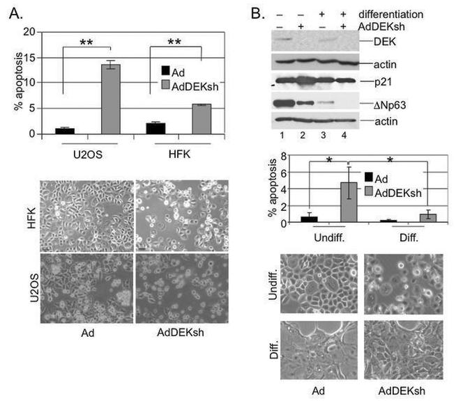
A. HFKs and U2OS cells were infected with either empty Ad or AdDEKsh and subjected to anti-active caspase 3 antibody and flow cytometry on day three. The cells were photographed after four days. B. HFK differentiation. HFKs were infected as in (A) and either subjected to normal medium, or differentiated upon the addition of 1mM calcium and 10% FBS. Cells were harvested for apoptosis assays as in A, subjected to DEK, p21, ΔNp63 and actin specific western blot analyses on day three, and analyzed for cellular morphology. One asterisk represents a p-value of < 0.05 while two asterisks represent a p-value of < 0.01. The Ad infected populations were not statistically different from each other nor were the Ad and DEKsh infected differentiated cell populations.