
Fig. 4. Animal testing of ICC scaffolds.
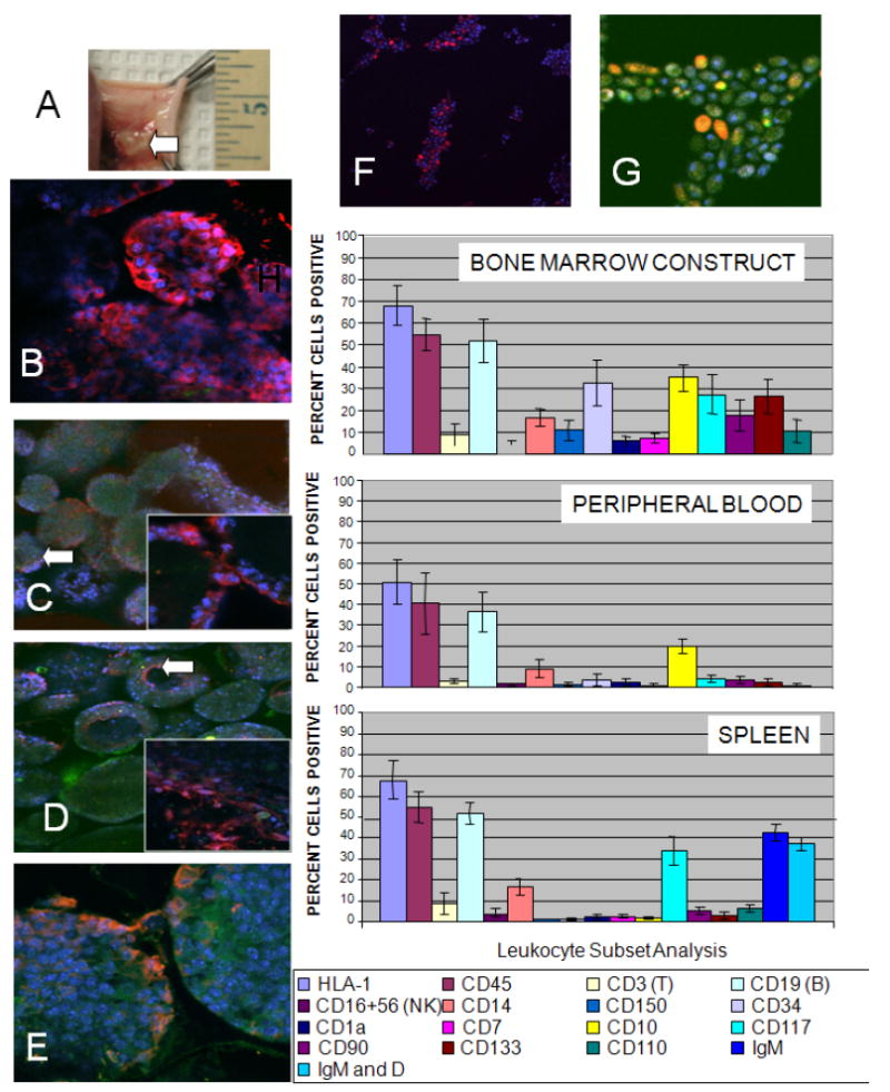
Evaluation of bone marrow construct, bone marrow derived cells, peripheral blood and spleen cells after 2 weeks implantation of hydrogel ICC scaffolds on the backs of eight SCID mice. The scaffolds were seeded with CFSE labeled cord blood derived CD34+ HSCs and cultured for 3 days before implantation. (A) A high degree of vascularization is seen in the regions near the site of the implanted ICC construct. (B-G) Confocal microscopy images of 7 μm frozen sections of the ICC-bone marrow construct. DAPI (blue) and CFSE (green). (B) Human MHC-Class I (red), 400X (green channel was not overlaid for clarity). HSC markers: (C) CD34 (red), insert 630X (D) CD150 (red), insert, 630X and (E) CD133 (red), (F) CD19 (red) (green channel not overlaid for clarity) and (G) IgM expression (red), magnification 630X. (H) Flow cytometry evaluation of cell phenotypes found in the bone marrow construct, peripheral blood and spleens of SCID mice receiving constructs. For all flow cytometry data 10,000 events were collected for each sample. Isotype control staining was less than 2% cells positive for all antibodies used.