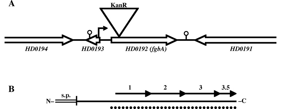
Fig. 1. Maps of the HD0192 (fgbA)-containing genetic locus (A) and the Fg-binding protein FgbA (B).
(A) Scale map of the fgbA locus in 35000HP. Open arrows indicate direction of transcription of ORFs; single lines indicate intergenic sequences. Triangle denotes site of insertional mutagenesis with KanR cassette in 35000HPfgbA. Small arrow indicates putative promoter upstream of the fgbA ORF; stalked circles indicate predicted Rho-independent transcriptional terminators. (B) Scale map of the protein product of fgbA. The horizontal solid line represents the 1° structure of the mature protein, the double line indicates the signal 2 peptide, and the vertical line between them denotes the site of signal peptide cleavage and lipidation. Arrows represent the 29-amino acid direct repeats. Dotted line indicates the predicted alpha-helical coiled-coil structure.

