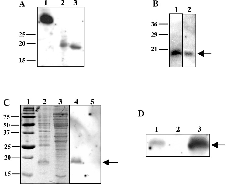
Fig. 2. FgbA conferred fibrinogen binding in a ligand blot assay.
(A) Lane 1, lysate of E. coli strain expressing recombinant Fg-binding portion of S. aureus ClfA; lane 2, lysate of E. coli strain expressing recombinant HD0192; lane 3, recombinant HD0192 after purification over a Ni column. (B) Whole cell lysate (lane 1) and OMP preparation (lane 2) of 35000HP probed with dig-Fg and developed to detect dig. Note the Fg-binding bands at 18 kDa (arrow). (C) Fg-binding activity was enriched in a hydrophobic protein preparation. Triton X-114 detergent (lanes 2, 4) or aqueous (lanes 3, 5) phase proteins from whole cell lysates of 35000HP were stained with Coomassie (lanes 2, 3) or probed with dig-Fg in the ligand blot (lanes 4, 5). Lane 1, molecular size markers. Note the 18 kDa bands (arrow) present in detergent phase samples. (D) FgbA conferred Fg-binding activity in H. ducreyi. Whole cell lysates of parent strain 35000HP (lane 1), mutant 350000HPfgbA (lane 2), and complemented mutant (lane 3) were probed with dig-Fg in the ligand blot assay. Note the FgbA band present in the parent, absent in the fgbA mutant, and restored in the complemented mutant.

