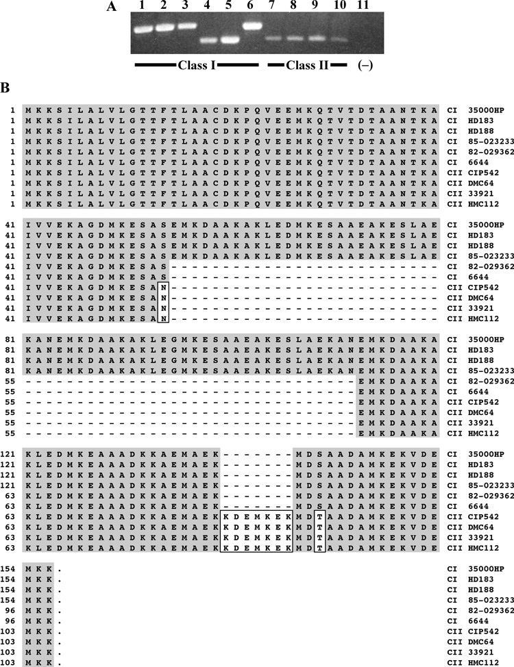
Fig. 3. fgbA varied in size but was conserved among class I and class II clinical isolates of H. ducreyi.
(A) PCR amplicons of the fgbA locus from genomic DNA of clinical isolates listed in Table 1. Lanes 1–6, class I strains 35000HP, HD183, HD188, 82-029362, 6644, and 85-023233; lanes 7–10, class II strains CIP542 ATCC, DMC64, 33921, and HMC112. Lane 11, negative control (no template added). (B) Alignment of deduced FgbA sequences among H. ducreyi clinical isolates. Strain names are listed on the right-hand side. C I, class I strain; C II, class II strain. Shaded residues match those of 35000HP, boxed residues differ from those of 35000HP, and dashes indicate missing residues.

