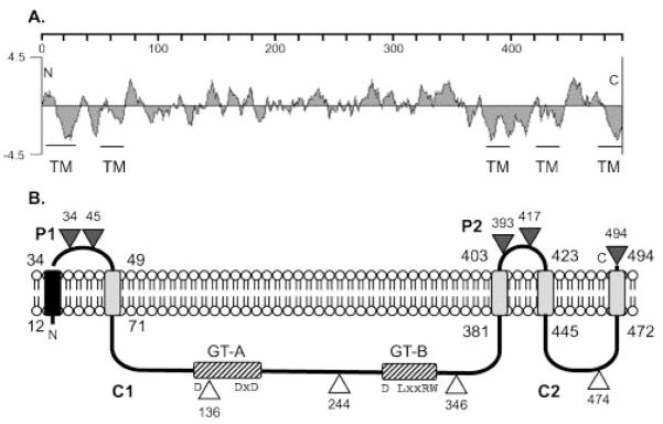
Fig. 1.
(A) A hydrophilicity plot of Alg8 and a TMpred analysis suggest five TM domains as indicated. The scale shows the residue numbers in Alg8. (B) Model for the orientation of Alg8 in the inner membrane based on the activities of in-frame Alg8′-PhoA fusions. Triangles show the fusion joints to PhoA, with the number showing the terminal Alg8 residue; black indicates positive activity (periplasmic) and white indicates negative activity (cytoplasmic). P1 and P2 are predicted periplasmic loops, and C1 and C2 are predicted cytoplasmic loops. The black rectangle in the membrane represents a TM domain predicted to be a cleavable signal peptide, and the grey rectangles represent other TM domains. The locations of GT domains A and B in cytoplasmic loop C1 are indicated with hatched rectangles. The widely spaced D, DxD, D, (L/Q)xxRW motif, highly conserved in the active site of family 2 GTs, is shown.

