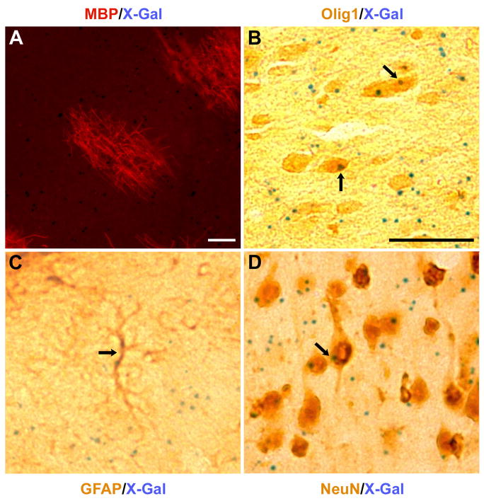
Fig. 5.
Dual immuno- and histochemical labeling of single sections, showing (A) an immunoreactive MBP patch along with punctate cellular X-gal reaction products from an “intermediate” chimeric mouse (MBP, red; X-gal, blue), and (B–D) co-localization of immunoreactive Olig1, GFAP, or NeuN (brown) with X-gal (blue, arrows) in presumptive oligodendrocytes (B), astroglia (C), and neurons (D) from a “rescued” chimeric mouse. Scale bars represent 50 μm.

