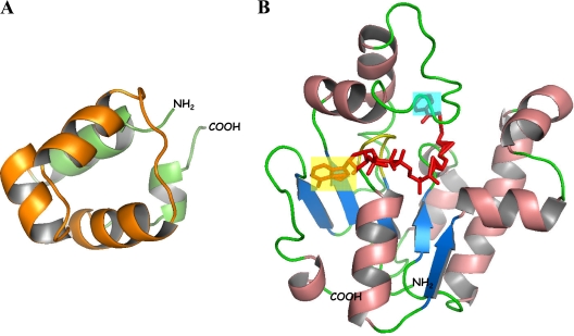
FIG. 21.
Three-dimensional model of the BzdR transcriptional regulator. (A) Ribbon diagram of N-BzdR showing the five-helix bundle. The helices and loop forming the classical HTH motif of XRE-type regulators are in orange. (B) Ribbon diagram of C-BzdR/benzoyl-CoA. The overall fold contains five β-strands (blue arrows) and eight α-helices (salmon ribbons). The phosphate binding site (P loop) is in yellow. Benzoyl-CoA is in red, with the benzoyl and ADP ends being blue and yellow shadowed, respectively. The corresponding N and C termini are labeled as NH2 and COOH, respectively. The figure has been prepared using MOLSCRIPT (197), RASTERED (243), and PyMOL (DeLano Scientific).

