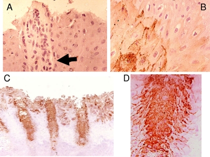
FIG. 2.
Immunohistochemical staining of treponemes in digital dermatitis lesions. (A) Staining of healthy hoof skin (hair follicle is identified by the arrow). (B) Staining of deep dermal layers showing treponemes tracking between cells. (C) Staining of hoof skin showing strong staining of hair follicles. (D) Staining of hair follicle showing tracking of infection from hair follicles into surrounding tissues.

