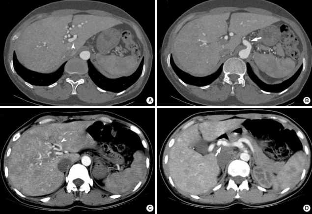
Fig. 3.
Radiology findings of the HHT patients. (A) and (B) CT angiography of the liver in patient 1 shows an enlarged celiac axis (arrow), a prominent hepatic artery (arrowhead) with multiple aberrant collateral vessels, and heterogeneous attenuation of the liver. (C) and (D) Abdominal CT of patient 3 shows severe tortuous dilatation of the hepatic artery (arrow) and its intrahepatic branches (arrowhead) with mottled hepatic enhancement.

