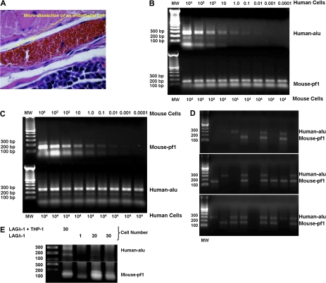
Figure 7.
Microdissection of single cells lining blood vessels from LAGλ-1 tumors containing THP-1 cells shows cells with the presence of 3 different types of DNA: only human, both human and mouse, and only murine sequences. (A) Single cells were microdissected from the lining of tumor blood vessels from mice injected with LAGλ-1 and THP-1 cells (100×/objective lens, Olympus BX51; Olympus). (B) To test the sensitivity to detect human DNA with the human-specific alu primers with PCR, we serially diluted human monocytes from 104 to less than a single cell with mouse liver cells. DNA was isolated with salmon sperm DNA protection and separated on 1% agarose gel following 40 cycles of PCR with human alu–specific or mouse pf1–specific primers. The results show that human-specific alu DNA can be detected to a sensitivity of 0.01 human cell equivalents. (C) Similarly, we determined the sensitivity to detect mouse-specific pf1 DNA by serially diluting mouse liver cells in human cells (U266 MM cell line). The results show that mouse-specific pf1 DNA can be detected to a sensitivity of 0.01 mouse cell equivalents. (D) Single cells were microdissected from tumor blood vessels derived from mice coinjected with LAGλ-1 and THP-1 cells, and DNA was isolated with salmon sperm DNA protection and separated on 1% agarose gel following 40 cycles of PCR with human alu–specific or mouse pf1–specific primers. Three different patterns of PCR-amplified products were identified: human DNA alone, mouse DNA alone, and both human and mouse DNA. (E) Single cells were microdissected from the lining of tumor blood vessels from mice injected subcutaneously with LAGλ-1 + THP-1 cells. DNA was isolated with salmon sperm DNA protection and separated on 1% agarose gel following 40 cycles of PCR with human alu–specific or mouse pf1–specific primers. The lanes containing 20 and 30 cells were derived from microdissected single cells that were combined together for PCR analysis. Single cells lining the tumor blood vessels derived from mice injected with human LAGλ-1 cells alone showed the presence of only mouse DNA in tumor blood vessels, whereas mice injected with LAGλ-1 and THP-1 cells together showed the presence of both human and mouse DNA.

