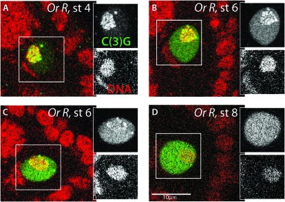
Figure 2.—
Developmental timing of SC disassembly. In the left panels DNA is red and C(3)G green; side panels show separated channels for boxed region, with C(3)G on the top. In wild-type oocytes C(3)G localizes to the meiotic chromosomes beginning in the germarium, prior to stage 2. This localization to the chromosomes, which are in a compact structure and take up only a small portion of the nucleus, is maintained through stage 4 or 5. As development continues, C(3)G begins to delocalize from the chromosomes and is seen with increasing intensity throughout the nucleus. (A–D) Display of the categories scored in Table 1. (A) C(3)G localized solely to chromosome axis (Oregon R, stage 4). (B) C(3)G dispersed on axis and spreading through nucleus (Oregon R, stage 6). (C) Trace amounts of C(3)G on chromosomes (Oregon R, stage 6). (D) No C(3)G associated with chromosomes (Oregon R, stage 8). All of the merged images are at the same magnification. Bar, 10μm.

