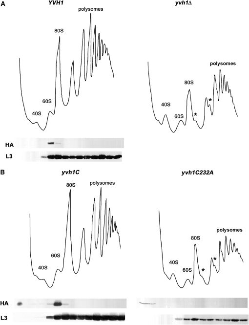
Figure 4.—
Yvh1 is associated with pre-60S ribosomes. (A and B) Ribosome fractionation. Cell lysate was prepared by vortexing with glass beads from the following strains: wild-type (KKY56) and yvh1Δ (KKY57) cells (A) or cells with yvh1-C terminus (KKY55) and full-length yvh1-C232A (KKY58) (B). Ribosomes were separated on 7–47% sucrose gradients and analyzed as described in materials and methods. Polysomes and 40S, 60S, and 80S ribosomes are indicated; asterisks indicate the positions of half-mer polysomes. Western blots show positions of HA-Yvh1 and the ribosomal protein L3 in gradients (bottom panels).

