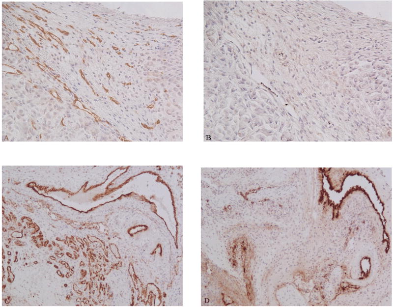
Fig. 5.
Comparision of anti-CD31 and anti-F VIII RAg stains using the optimized AR method. Serial sections from MDA-MB-231 breast cancer xenograft (A and B) and syngeneic breast cancer (C and D). Sections were treated with 0.5 M Tris buffer (pH 10) followed by the CD31 immunohistochemistry (A and C). Sections were treated with 0.05% pepsin followed by F VIII RAg immunohistochemistry (B and D). Anti-CD31 produced better microvessel staining compared to anti-F VIII RAg staining. A and B: 200 X; C and D 100 X.

