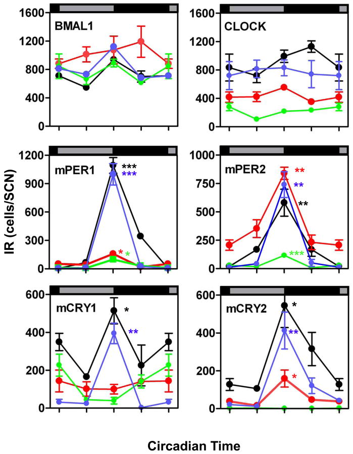
Figure 4. Number of cells with BMAL1-, CLOCK-, mPER1-, mPER2-, mCRY1-, and mCRY2-immunoreactive cells in the intermediate portion of the SCN in fetal (green), newborn (red), infantile (blue), and adult (black) mice.
Data are expressed as the mean +/− SEM of five animals per timepoint and ontogenetic stage. Data points at CT 00/24 are double-plotted. Asterisks indicate significant differences between peak values at CT12 and trough values at CT00 (*=p<0.05, **=p<0.01, ***=p<0.001; one-way ANOVA).

