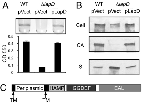
Fig. 1.
Biofilm formation and LapA localization phenotypes of the lapD mutant. (A) Quantitative analysis of biofilm formation by WT plus vector (WT pvect), ΔlapD plus vector (pvect), and ΔlapD plus pLapD (pLapD). (B) Western blots probed for LapA to analyze adhesin localization profiles for strains shown in (A). The fractions indicated are cellular (Cell), cell-associated (CA), and culture supernatant (S). (C) Predicted protein domains of LapD.

