Fig. 4.
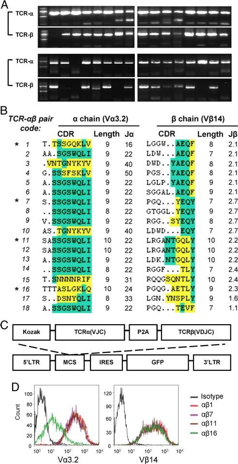
TCR Cloning. (A) Every TCR pair including the full-length α chain and the full-length of β chain was PCR amplified from the same single CD4+Vα3.2+Vβ14+ FYAK-specific T cell. PCR products from 32 single cells are shown. Only those pairs with good amplification of both α and β chain were used for further subcloning. (B) CDR3 amino acid sequences for Vα3.2 and Vβ14 TCR from 18 individual cells. The conserved residues CAXS or CAWS flanking the 5′ CDR3 of Vα3.2 and Vβ14 respectively, or FGXG flanking the 3′ CDR3 is not shown. Residues found in the J region are indicated in green. The amino acids different from the most frequent residues of the J region are indicated in yellow. The asterisk denotes the 4 TCR αβ pairs used for subcloning. (C) Structure of retroviral TCR construct. Amplified TCR α and β chain cDNA sequences were linked with the 2A peptide sequence of porcine teschovirus-1 (P2A) by recombinant PCR and then subcloned into a cassette vector, MSCV-IRES-GFP (pMIG), for retrovirus-mediated expression. (D) 293T cells cotransfected with TCR construct and CD3 vector (pMIG-CD3δγεζ) were stained with mAbs against TCR Vα3.2 and Vβ14. Flow cytometry showing the expression of cloned TCR on the surface of transfected 293T cells.

