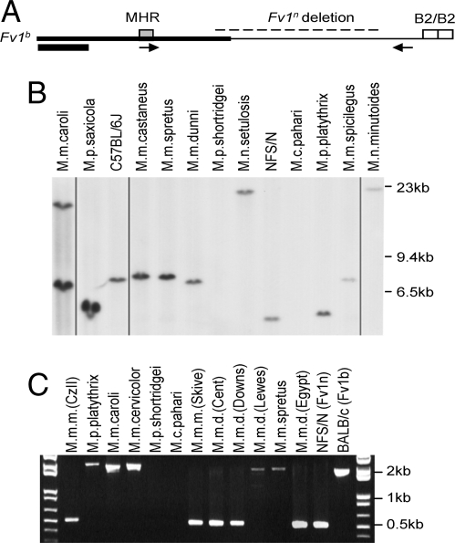
Fig. 1.
Detection of Fv1 in DNAs of Mus species. (A) The structure of Fv1b is shown with a gray box marking the MHR, open boxes representing B2 repeats and a dashed line representing the 1.3-kb segment deleted in Fv1n. The arrows represent the PCR primers Fb3003 and Rb4831, and the black box represents the segment used for blot hybridization. Fv1 is encoded by a single exon. (B) Southern blot analysis of BglI-digested Mus DNAs. All lanes taken from the same exposure of a single blot; deleted lanes are indicated by vertical lines. (C) PCR products of Mus DNAs.

