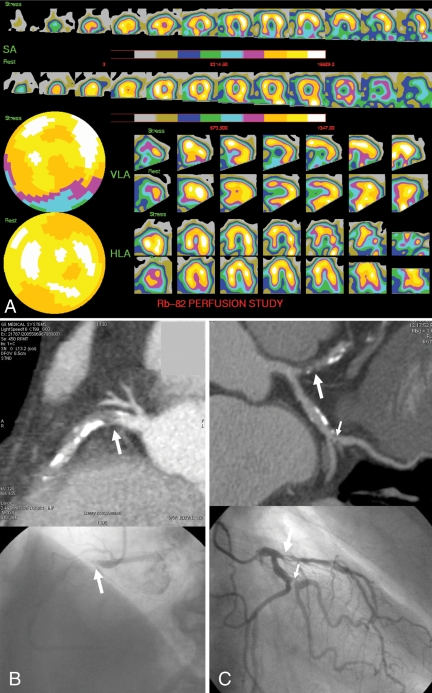
Figure 1).
Positron emission tomography magnetic perfusion imaging, computed tomographic angiography and conventional invasive coronary angiography of a 73-year-old man with chest pain. A Positron emission tomography myocardial perfusion imaging: post-stress, a moderate (blue) reduction in rubidium-82 (Rb-82) uptake is present in the basal and mid-inferior wall, with normalization at rest. B Curved multiplanar reformation computed tomography and conventional invasive coronary angiography images demonstrating an occluded proximal right coronary artery (arrow). C Significant stenosis of the proximal left anterior descending coronary artery (large arrow) and the first marginal artery (small arrow). HLA Horizontal long axis; SA Short axis; VLA Vertical long axis

