Fig. 4.
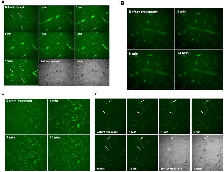
Protein extracts from S. macrurus blastema (SmBE) induced calcium transient in myotubes. Differentiated C2C12 cells were loaded with calcium indicator Fluo-4 for 1 hr before additional treatment for the indicated time periods. Time-lapse pictures were taken every minute. A: Representative time lapse images of Fluo-4 signal after addition of SmBE to the culture during 15-min period. The fluorescence signal was detected at excitation 485 nm and emission at 520 nm wavelength, and time-lapse images were taken at ×100 magnification. B: Representative time lapse images of Fluo-4 signal after addition of adult muscle extract to the culture during a 14-min period. C: Calcium chloride (20 mM) was added to C2C12 cells loaded with fluo-4. Representative time lapse images of 10 min period are shown. D: Caffeine (40 mM) was added to the culture of C2C12 cells loaded with fluo-4 and time lapse images were taken for 15-min period every minute. Representative images are shown. Arrows point individual myotubes that contracted after calcium transient. Images were taken at ×100 magnification.

