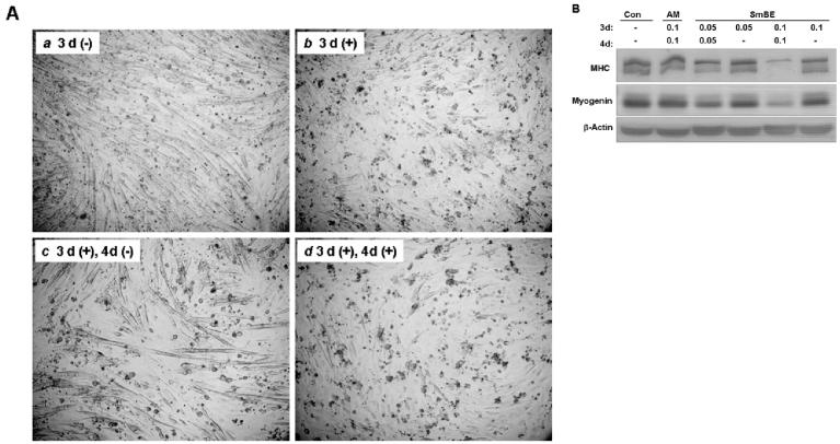
Fig. 8.
Inhibition of myoblast differentiation by Protein extracts from S. macrurus blastema (SmBE) is reversible. A: C2C12 myoblasts were cultured in differentiation medium (DM) only (a), or in DM with SmBE (b) for 3 days. Myoblasts treated with SmBE (0.1 mg/ml) for 3 days (b) were replaced with DM only and incubated for 4 additional days (c). To compare the effect of SmBE removal from DM, myoblasts were continued to be incubated in DM with SmBE for 4 additional days (d), in parallel with (c). Phase contrast images were taken at ×100 magnification. 3d(−): DM only for 3 days; 3d(+): DM and SmBE for 3 days; 3d(+), 4d(−): DM and SmBE for 3 days followed by DM only for 4 days; 3d(+), 4d(+): DM and SmBE for 7 days. B: Western blots of C2C12 myoblasts cultured in different conditions for 7 days using antibodies against sarcomeric MHC, myogenin and β-actin. Total protein (50 μg) extracted from myoblasts was resolved by 4-15% sodium dodecyl sulfate-polyacrylamide gel electrophoresis, transferred to a polyvinylidene difluoride (PVDF) membrane, and probed with antibodies. β-actin immunoblot was done to demonstrate equal loading. Con, DM only; AM, adult muscle extract; SmBE, S. macrurus blastema extract. Concentrations of protein extract were 0.05 mg/ml and 0.10 mg/ml.

