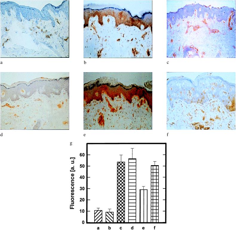
Figure 2.
Restoration of UVB radiation-induced inhibition of IFN-γ-induced keratinocyte ICAM-1 expression by photoreactivation. (a–f) Keratinocyte ICAM-1 surface expression in human buttock skin that had been left untreated (a); i.c. injected with rh IFN-γ (b); UVB-irradiated (1 MED) (c); UVB-irradiated and subsequently i.c. injected with rh IFN-γ (d); UVB-irradiated, then topically treated with photolyase-containing liposomes, followed by exposure to photoreactivating light and subsequent i.c. injection with rh IFN-γ (e); or UVB-irradiated, then exposed to photoreactivating light, followed by topical treatment with photolyase-containing liposomes and subsequent i.c. injection with rh IFN-γ (f). ICAM-1 surface expression was detected by immunohistochemistry with mAb 84H10 as described in Materials and Methods. Data represent one of three essentially identical experiments. (g) Semiquantitative analysis of epidermal pyrimidine dimer content in skin areas a–f. The presence of pyrimidine dimers was assessed 22.5 h after photoreactivation and quantified by immunfluorescence microscopy with mAb H3 as described in Materials and Methods. Data are given as histograms of specific fluorescence in arbitrary units (a. u.) as described (16) versus skin area and represent mean values ± SD of three experiments.

