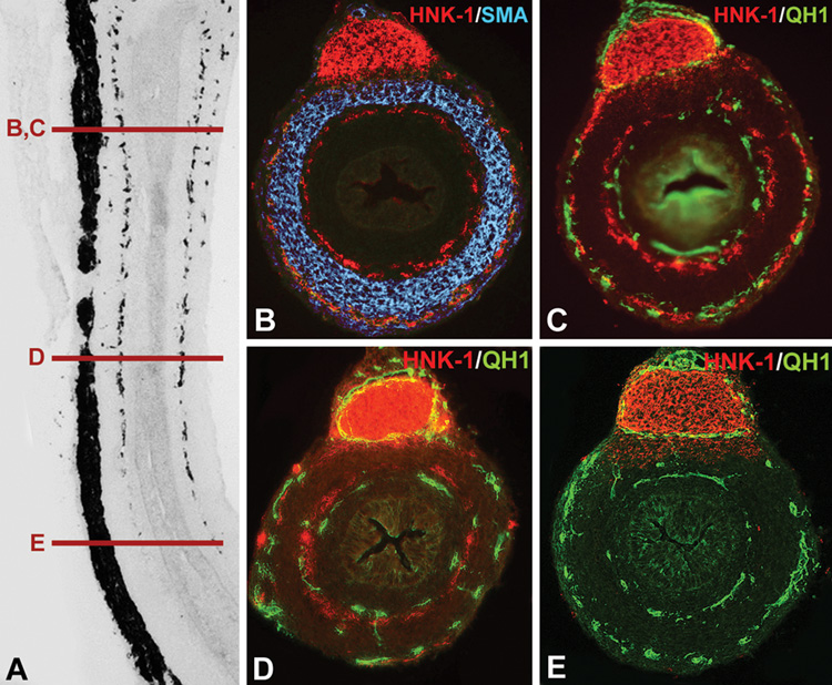
Figure 3. Endothelial cells presage ENCC patterning in the avian embryonic gut.
A longitudinal section of an E7 quail colorectum was labeled with the neuronal antibody, Hu (A), and multiple cross-sections taken at the levels marked (B–E). In a proximal section (B), two plexuses of ENCCs are seen on either side of the circular muscle, labeled with smooth muscle actin (SMA). Double-labeling with the endothelial cell marker, QH1, and the neural crest cell marker, HNK-1, shows that ENCCs are positioned adjacent to endothelial cells (C). In a more distal section through the mid-colorectum, ENCCs are present mostly in the submucosal plexus, with fewer cells in the myenteric plexus, while two rings of endothelial cells are present (D). In the distal colorectum, where ENCCs have not yet colonized, endothelial cells are already patterned into two concentric rings (E).

