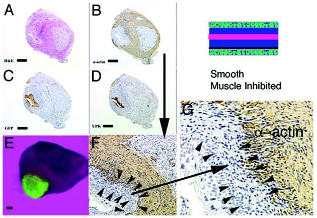
Figure 5.

Schematic and results of urothelial recombination with an intact bladder in which ectopic urothelium was also placed in the serosal location, and thus both orthotopic and heterotypic urothelia were present. Histologic serial sections: (A) H&E = hematoxylin and eosin; Immunohistochemistry: (B) α-actin = smooth muscle alpha actin; (C) GFP=Green Fluorescent Protein; (D) UPK= uroplakin; (E) Gross image of the GFP-positive graft; (F) higher magnification (3X) of (B, α-actin) showing two zones devoid of smooth muscle (between arrowheads and also large arrow); (G) higher magnification (2X) of (F) showing urothelium, zone devoid of smooth muscle and α-actin-positive smooth muscle. Note the zone of smooth muscle inhibition between the serosal ectopic urothelium and smooth muscle (between arrowheads). (magnification bar = 100um)
