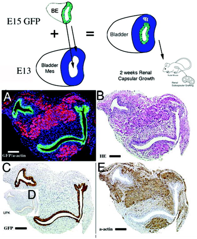
Figure 6.

Schematic and results of urothelial recombination with bladder mesenchyme in heterotypic and orthotopic locations. Histologic serial sections: (A) Color fluorescent triple stain, GFP is green, alpha-actin is pink and Hoescht dye is blue representing the zone of smooth muscle inhibition or submucosa around the placed urothelium; (B) H&E = hematoxylin and eosin; Immunohistochemistry: (C) GFP=Green Fluorescent Protein; (D) UPK= uroplakin and (E) α-actin = smooth muscle alpha actin. (magnification bar = 100um)
