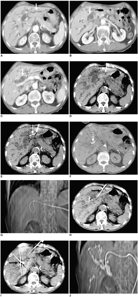
Fig. 1.
65-year-old male patient with distal CBD cancer.
A-C. CT scan images show dilated intrahepatic and extrahepatic ducts. It is possible to trace advancement course of guide wire and catheter on CT images (white arrows).
D-F. Monitoring CT fluoroscopy images, segment 3 IHD is punctured and guide wire is subsequently inserted, finally to CBD (dotty arrows).
G. Tip of guide wire is confirmed to be placed in CBD on scanography image.
H, I. Drainage catheter is inserted over guide wire monitoring CT fluoroscopy images (arrows).
J. Cholangiography image is acquired using scanography after finishing procedure.

